He Described Microbial Cells Using a Compound Microscope
The highest resolution possible is around 02 μm. The invention of the microscope led to the discovery of the cell by Hooke.
The Microscopic World Microbiology A Laboratory Experience
Marcello Marpighi known as the father of microscopic anatomy found taste buds and red blood cells.

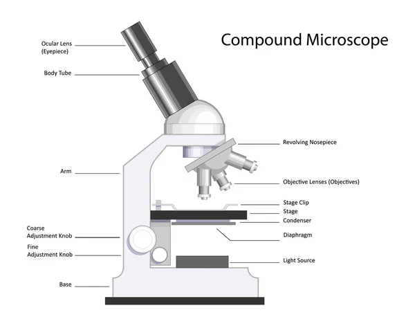
. The _____ lens is found in the eyepiece and the _____ lens is found in the revolving nosepiece. Simple microscopes have a single lens while compound microscopes have multiple lenses. Antony van Leeuwenhoek was born on October 24 1632 in Delft Netherlands.
Bottom part of the microscope stabilizes it and allows it to stand upright. He described a new world alongside exquisite drawings of the stinging hairs on a nettle a flea and most famously of all the honeycomb structure or cells of a cork. This discovery led to the development of the classical cell theory.
Interestingly while Hooke did use a compound microscope he found that it much strained and weakened his sight. He was the first to coin the name cell after he observed the angular spaces that he saw in a thin section of cork. The term compound in compound microscopes refers to the microscope having more than one lens.
Antonie van Leeuwenhoek is credited with the first observation of microbes including protists and bacteria with simple microscopes that he made. - affix the cells to the slide. With the microscope you will be able to observe.
All basic chemical physiological functions are carried out inside the cell movement digestion etc 7. A compound microscope uses two lenses at once to magnify the image of a specimen. Simple microscopes have a single lens while compound microscopes have multiple lenses.
There are more than two lenses in a compound microscope. While they are very small they are diverse and vary in shape and size. Robert Hooke was the first to describe what we now call cells.
Learn about the working principle parts and uses of a compound microscope along with a labeled diagram here. The maximum total magnification for a microscope using visual light for illumination is around 1500X where the microscope might have 15x oculars and a 100x oil immersion objective. If objects or cells are closer together than this they cant be distinguished as separate entities.
The correct answer is A. 6 Post-Lab Questions 1. It is U or horseshoe-shaped metallic structure.
Like archeans bacteria are prokaryotic cells. He used a simple microscope. What discovered the microscope.
All cells are basically the same in chemical composition and metabolic activities. Cell activity depends on the activities of sub-cellular structures within the cell organelles nucleus plasma membrane 3. Robert Hooke publishes Micrographia and reveals his refinements to the compound microscope enabled him to describe cells molds plants and insect parts such as compound eyes of flies small parasites such as lice and fleas.
Bacteria Under the Microscope Types Morphology and Reproduction. Devised with a system of combination of lenses a compound microscope. Robert Hooke was the first to describe what we now call cells.
These ideas were formulated by a number of early biologists. Anton van Leeuwenhoek can be easily described as the most unlikely person ever to contribute to the world of science. Human blood cells are larger than Bacillus subtilis.
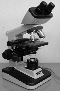
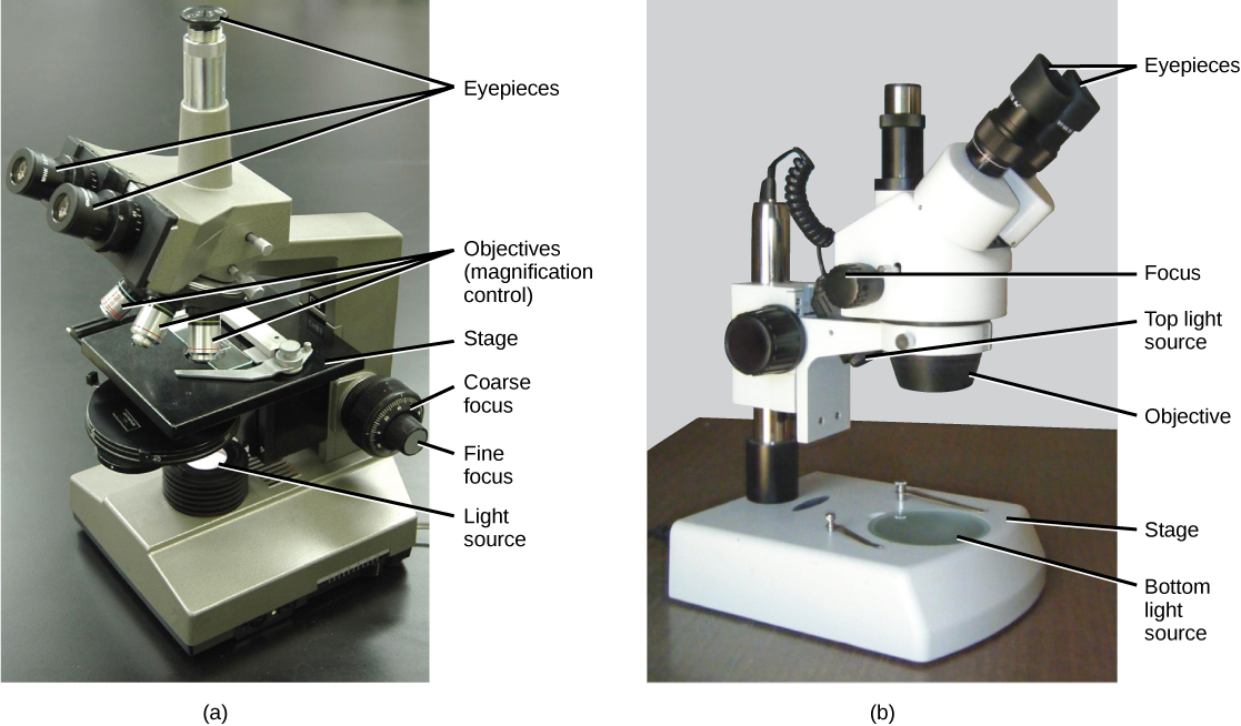
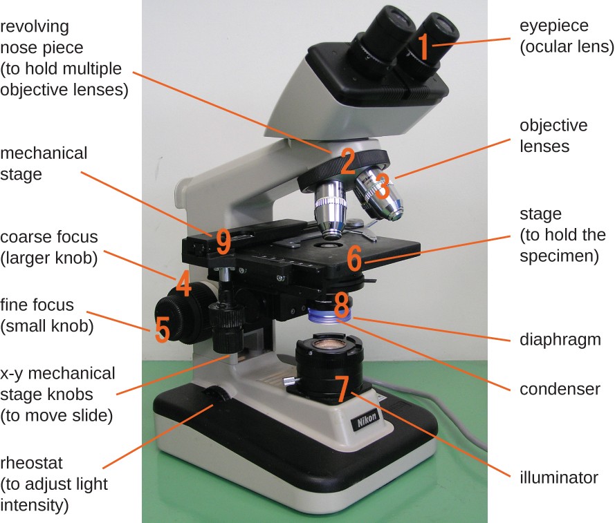
Yeast are single-celled eukaryotes that are smaller than blood cells but much larger than prokaryotes such as Escherichia coli. Proper Use of the Compound Light Microscope Exercise 1B Steps to follow when using the microscope If you really want to be able to see a specimen on a slide you must follow the steps on the next page every time you look at a new slide. A modern compound microscope has following structural components.
This means that they are single-celled organisms without a nucleus membrane nuclear envelope. The microscope will be your friend if you always use the following steps in their proper order. Compound microscope is a type of optical microscope that is used for obtaining a high-resolution image.
Describe how you can quantify the size difference between the tick and the mite using the microscope. The optical microscope often referred to as the light microscope is a type of microscope that uses visible light and a system of lenses to magnify images of small subjects. The Cell Theory The idea that cells are fundamental units of life is part of the cell theory.
A typical animal cell is 1020 μm in diameter which is about one-fifth the size of the smallest particle visible to the naked eye. It is used for passive observation of structural details of a cell tissue or organ in sections. A compound microscope is an indispensable instrument in any biological laboratory.
This discovery proposed as the cell doctrine by Schleiden and. Antonie van Leeuwenhoek is credited with the first observation of microbes including protists and bacteria with simple microscopes that he made. Robert Hooke discovered cells by studying the honeycomb structure of a cork under a microscope.
40 Which microscope is best used for observing the surfaces of intact cells and viruses. Connects the base to the Binocular Tube. Why must specimens viewed with a compound microscope be thin or chemically cleared.
Which of the following statements is TRUE regarding the relative sizes of the different individual cells. Where was the first microscope invented. He described the use of the compound microscope that he had devised.
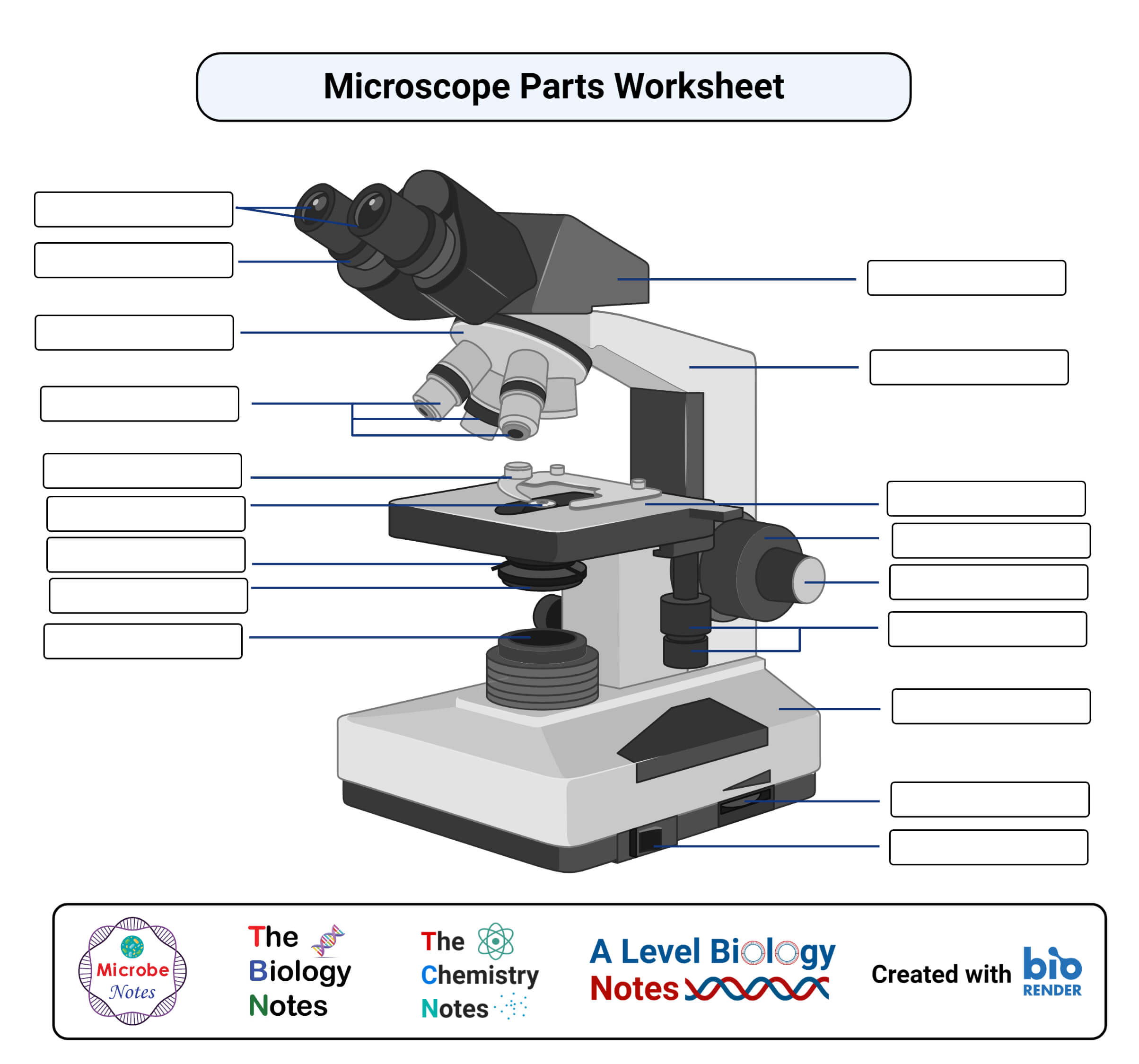
While looking at cork Hooke observed box-shaped structures which he called cells as they reminded him of the cells or rooms in monasteries. Parts of a Compound Light Microscope. 42 The resolution of a microscope can be improved by changing the -wavelength of light.
41 Bacterial smears are fixed before staining to -affix the cells to the slide. Robert Koch used a compound microscope to discover tubercle and cholera bacilli. Unlike van Leeuwenhoeks single lens microscope we now combine the magnifying power of multiple lenses in what is called a compound microscope.
In this lab you will use the compound light microscope to observe these same kinds of cells. It was not until good light microscopes became available in the early part of the nineteenth century that all plant and animal tissues were discovered to be aggregates of individual cells. It was Hooke who coined the term cells when describing living tissue.
The mite is significantly smaller than the tick. It has to be thin so the light can make it through the object allowing it to be visible in the compound microscope. Was the first to observe human blood cells and to observe yeast and bacteria which he referred to as animalcules.

Different Parts And Functions Of Compound Microscope

Lab Manual Exercise 1

What Is A Compound Microscope Microscope World Blog

Compound Microscope Definition Labeled Diagram Principle Parts Uses
3 1 How Cells Are Studied Concepts Of Biology 1st Canadian Edition
Instruments Of Microscopy Microbiology

Techniques Using A Microscope To Explore Fermented Foods Microbialfoods Org

Cell Morphology Under Compound Microscope Download Scientific Diagram
Parts Of A Microscope With Functions And Labeled Diagram

Compound Microscope Parts Diagram Definition Application Working Principle

Ch 2 Homework Flashcards Quizlet
337 Microscope Slide Vector Images Microscope Slide Illustrations Depositphotos
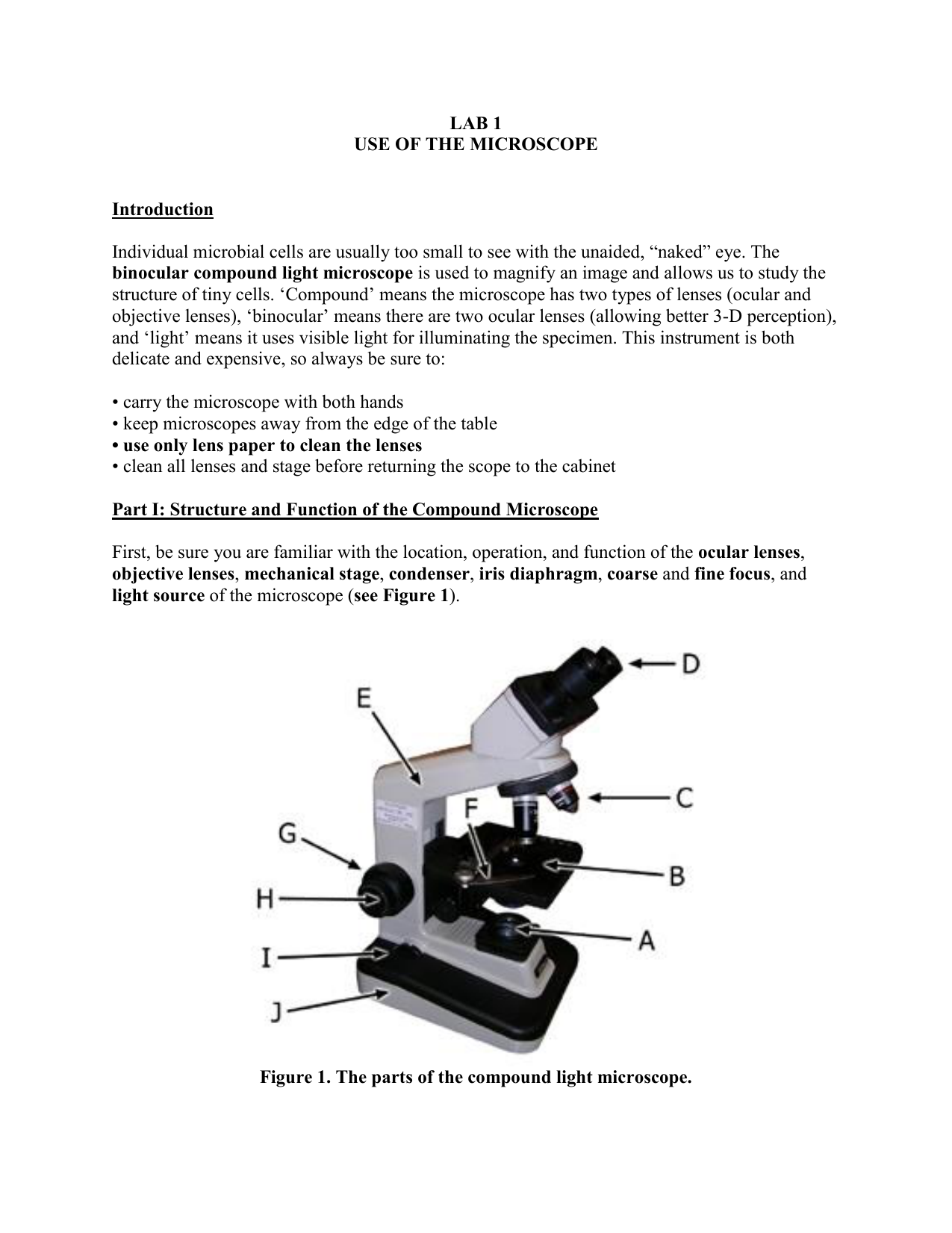
Lab 1 Use Of The Microscope Introduction Individual Microbial Manualzz

Applications Asbestos Water Soil Environmental Microscopes Soil Tr Amscope

What Is A Compound Microscope Microscope World Blog

Introduction To The Oil Immersion Compound Microscope Ppt Download

Figures Show The Morphological Characteristics Of Bacterial Isolated Download Table

Observing Bacteria Under The Light Microscope Microbehunter Microscopy

Compound Microscope Definition Diagram Parts Uses Working Principle


Comments
Post a Comment